


Coxartrosis- this is the arthrosis of the hip union.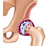 It develops gradually, for several years, prone to progress, can be both unilateral and double.It is associated with pain and restriction of movements in the union.In the later stages, hip muscle atrophy and shortening of the limbs are observed.The diagnosis is established on the basis of clinical symptoms and radiography results.In the early stages of coxarthrosis, conservative treatment.With the destruction of the joint, especially in young and middle -aged patients, surgery is indicated.
General information
Coxarthrosis (osteoarthrosis or deforming arthrosis of the hip joint) is a degenerative-degenerative disease.It usually develops at the age of 40 and older.It can be the result of various damage and common diseases.Sometimes it happens without any obvious reason.Coxarthrosis is characterized by a gradual progressive course.In the early stages, conservative treatment methods are used.In later stages, the common function can only be restored operational.
In orthopedics and traumatology, coxarthrosis is one of the most common arthrosis.The high frequency of its development is due to a considerable load on the hip node and the widespread spread of congenital pathology - joint dysplasia.Women suffer from coxartrosis slightly more often than men.
Causes of Coxartrosis
The main arthrosis (arising for unknown reasons) and secondary (developed as a result of other diseases) of the hip joint are distinguished.
Secondary coxetrosis may be the result of the following diseases:
- The joint dysplasia of the hip.
- The innate displacement of the thigh.
- Diseases reach.
- Aseptic necrosis of the thigh head.
- Infectious lesions and inflammatory processes (for example, hip joint arthritis).
- Injuries (traumatic displacements, hip neck fractures, pelvic fractures).
Coxarthrosis can be either one -sided or double.With primary coxarthrosis, a simultaneous lesion of the spine (osteochondrosis) and knee (gonartrosis) is often observed.
Risk factors
Among the factors that increase the chance of developing coxarthrosis include:
- Continuous increased load on coupling.It is most often observed in athletes in people with excess body weight.
- Disordishes circulatory disorders, hormonal changes, metabolic disorders.
- Pathology of the spine (kyphosis, scoliosis) or stop (flat feet).
- Elderly and old age.
- A sedentary lifestyle.
Coxarthrosis itself is not inherited.However, certain features (metabolic disorders, structural traits of the skeleton and weakness of the cartilage) can be inherited by the child by parents.Therefore, in the presence of blood relatives suffering from coxarthrosis, the probability of the onset of the disease has increased slightly.
Patanatomy
The hip connection is formed by two bones: ileum and femoral.The thigh head is articulated with the iliac bone acetabulum, forming a special "hinge".During the movements, acetabulum remains stationary, and the femur's head moves in different directions, providing flexion, stretching, abduction, behavior and rotating buttocks.
During the movements, the articular surfaces of the bone slides unhindered compared to one another, thanks to the smooth, elastic and durable cartilage of the hyaline covering the reversible cavity and the thigh head.In addition, the Hialin cartilage performs a function of overturning the shock and is involved in the redistribution of the load while moving and walking.
In the joint cavity there is a small amount of joint fluid, which plays the role of lubrication and provides the food of the hyaline cartilage.The connection is surrounded by a dense and strong capsule.Above the capsule are the large femur and gluteal muscles, which provide movements in the joint and, along with the hyaline cartilage, are also shock absorbers that protect the joints from unsuccessful movement damage.
With coxarthrosis, the joint fluid becomes thicker and more viscous.The surface of the hyaline cartilage dries, loses softness, covered with cracks.Due to the rudeness that has arisen, the cartilage during movements is constantly damaged for each other, which causes them to thin and worsen the pathological changes in the union.As coxarthrosis progresses, the bones begin to deform, "adaptation" to added pressure.Metabolism in unity is deteriorating.In the later stages of coxarthrosis, the severe atrophy of the injured limb muscles is observed.
Symptoms of coxarthrosis
The main symptoms of the disease include pain in the joint, the inguinal region, the thigh and the knee joint.Also, with cokesarthrosis, stiffness of movements and stiffness of the joint, walking disturbance, dementia, hip muscle atrophy and shortening of the limbs on the lesion side are observed.A characteristic feature of coxartrosis is a abduction restriction (for example, the patient is difficult when trying to sit in a chair).The presence of certain signs and their severity depends on the stage of the coxarthrosis.The first and most persistent symptom is pain.
onFirst degree coxetrosisPatients complain of periodic pain, which occurs after physical activity (prolonged running or walking).The pain is located in the joints, less often in the thigh or knee.After rest, it usually disappears.The strolling for 1 degree coxarthrosis does not break down, movements are completely stored, no muscle atrophy.
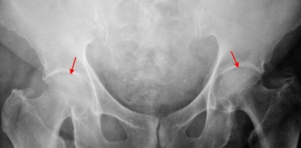
In the X -ray of the patient suffering from the 1 degree coxarthrosis, mild changes are determined: the uneven moderate narrowing of the joint gap, as well as the bone growth around the outer or inner edge of the acetabule in the absence of femur's head and neck changes.
toCoxartrosis 2 degreesThe pain becomes stronger, often appears at rest, radiates to the thighs and groin.After a significant physical activity, the patient with coxartrosis begins to lame.The volume of movements in the union decreases: the internal abduction and rotation of the thigh is limited.
In the X -Ray images for the 2nd degree coxarthrosis, the important uneven narrowing of the joint gap (more than half of the normal height) is determined.The femur's head is displaced somewhat up, deformed and increases in size, and its contours become uneven.Bone growths with this degree of coxarthrosis appear not only in the interior but also on the outer edge of the acetabulum and go out of the cartilage.
toCoxartrosis 3 degreesThe pain becomes persistent, the concern of patients not only during the day but also at night.Walking is difficult when moving, a patient with coxarthrosis is forced to use a cane.The volume of movements in the joint is severely limited, the muscles of the buttocks, buttocks and lower legs are atrophied.The weakness of the thigh muscles of the thigh causes the pelvic deviation to the anterior plane and the shortening of the limbs on the sore side.To compensate for the abbreviation, a patient suffering from coxarthrosis, when walking, bends the body in the injured direction.Because of this, the center of gravity is displaced, the load on the injured joint increases significantly.
In radiographs for 3rd degree coxarthrosis, a sharp narrowing of the joint gap is detected, a pronounced expansion of the thigh head and numerous bone growth.
Troubleshooting
The diagnosis of coxarthrosis is based on clinical signs and data of additional studies, the main of which is radiography.In many cases, the X -rays make it possible to place not only the degree of coxarthrosis, but also the cause of its appearance.Thus, for example, an increase in the angle of the diaphysis in the neck, scenes and flattening of acetabulum indicate dysplasia, and changes in the form of the proximal femur part are shown that coxarthrosis is a consequence of God's disease or youth epiphysiolis.In radiographs of patients with coxarthrosis, changes can also be detected by indicating injuries.
How can other methods of instrumental diagnosis of coxarthrosis, CT and MRI be used.The calculated tomography allows you to study in detail the pathological changes from the detailed bone structures, and the image of magnetic resonance offers the opportunity to evaluate soft tissue disorders.
Diagnosis
First of all, coxarthrosis should be differentiated from gonarthrosis (knee osteoarthrosis) and osteochondrosis of the spine.Muscle atrophy, which occurs in 2 and 3 stages of coxarthrosis, can cause pain in the knee joint, which are often expressed brighter than pain in the damage area.Therefore, with the patient's complaints about knee pain, a clinical (inspection, palpation, volume determination of movements) is the study of the hip node, and if coxarthrosis is suspected, to direct the patient to radiographs.
Pain for radical syndrome (nerve root compression) for osteochondrosis and some other spinal diseases can mimic pain with coxarthrosis.Unlike the coksartrosis, when squeezing the roots, the pain appears suddenly, after an unsuccessful movement, a sharp twist, the weight of the lift, etc., localizes to the area of the buttocks and spreads along the back of the thigh.A positive symptom of tension is detected - severe pain when the patient tries to raise a directed limb, lying on his back.At the same time, the patient takes the leg freely on the side, while in patients with coxarthrosis, the abduction is limited.It should be borne in mind that osteochondrosis and coxartrosis can be observed at the same time, therefore, in all cases, a thorough examination of the patient is needed.
In addition, the cokesarthrosis is differentiated with trochantheritis (Boot buzz) - aseptic inflammation in the area of gluteal muscle binding.Unlike coxarthrosis, the disease develops rapidly, within 1-2 weeks, usually after an injury or significant physical activity.The intensity of the pain is higher than with coxartrosis.Movement restrictions and limb cuts have not been observed.
In some cases, with an atypical course of the disease or reactive arthritis, symptoms that resemble coxarthrosis may be observed.Unlike coxarthrosis, with these diseases, the peak of pain falls at night.Pain syndrome is very intense, it can decrease when you walk.Breakfast stiffness is characteristic, which occurs immediately after you wake up and gradually disappears within hours.
Treatment of coxarthrosis
Pathology treatment is engaged in traumatologist orthopedists.The choice of treatment methods depends on the symptoms and stage of the disease.In stages 1 and 2 of coxarthrosis, conservative therapy is performed.During the period of irritation of coxarthrosis, injection blocks, non -steroidal anti -inflammatory drugs (pyroxes, indomethacin, diclofenac, ibuprofen, etc.).It should be borne in mind that medicines of this group are not recommended for a long time, as they can have a negative effect on the internal organs and suppress the ability of the hyaline cartilage to restore.
To restore the damaged cartilage for coxartrosis, funds are used by a group of chondroprotectors (chondroit sulfate, cartilage extract, etc.) are used.To improve blood circulation and eliminate the spasm of small vessels, vasodilating drugs (zinnaritis, nicotine acid, pentoxifillin, nicotine xanthinol) have been prescribed.According to indications, muscle relaxants (relaxing muscle drugs) are used.
With stubborn pain syndrome, patients suffering from coxarthrosis may be prescribed intra -articular injections using hormonal medicines (hydrocortisone, triamcinolone, metrumor).Treatment with steroids should be done carefully.Moreover, with coxarthrosis, local products are used - heat drives that do not have a pronounced therapeutic effect, however, in some cases they relieve muscle spasm and reduce the pain due to their "distinctive" action.Also, with coxarthrosis, physiotherapeutic procedures are prescribed (Excellent therapy, ultrasonic, laser treatment, UHF, induction, magnetotherapy), massage, manual therapy and therapeutic gymnastics.
The diet for coxarthrosis does not have an independent therapeutic effect and is used only as a tool to reduce weight.Lowering body weight allows you to reduce the load on the hip joints and, as a result, to ease the flow of coksartrosis.To reduce the load on the joint, the doctor, depending on the degree of coxarthrosis, may recommend walking with a cane or crutch.
In the later stages (with 3th degree coxarthrosis), the only effective method of treatment is surgery - replacing the destroyed node with an endoprostese.Depending on the nature of the lesion, it can be used either with a single pole (replacing only the thigh head) or two -pole (replacing both the thigh head and the reversible cavity).
The functioning of the endoprosthetics for coxarthrosis is performed in a planned manner, after a thorough examination, under general anesthesia.In the post -surgery period, antibiotic therapy is performed.The dresses are removed in 10-12 days, after which the patient is prescribed for outpatient treatment.After the endoprosthetics, rehabilitation measures are necessarily kept.
In 95% of cases, surgical intervention to replace joining with coxarthrosis ensures complete restoration of limb function.Patients can work, move actively and even play sports.The average service life of the prosthesis, undergoing all recommendations, is 15-20 years.After that, a second surgery is required to replace a worn endoprostese.